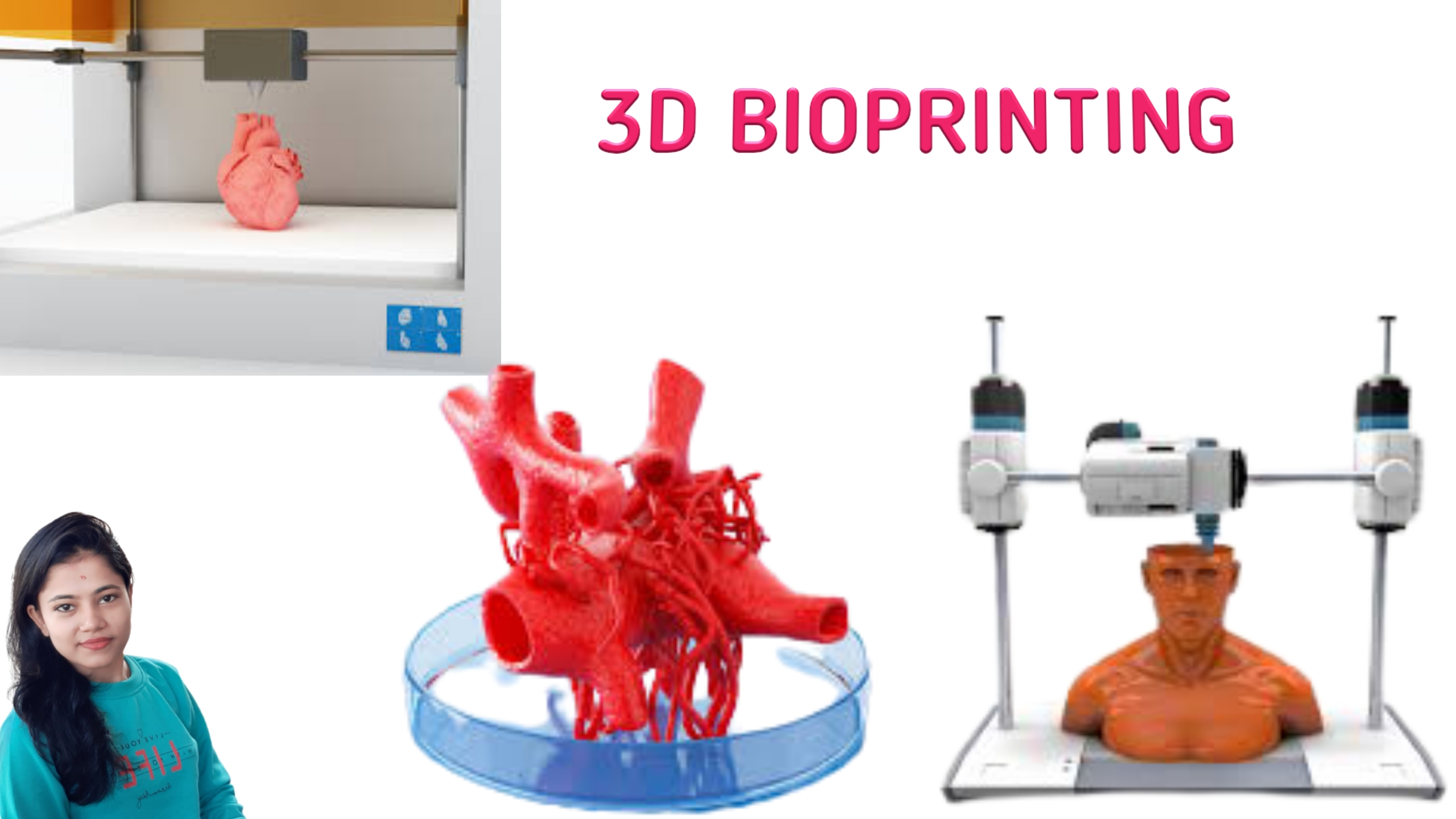
In this article, you know about 3D Bioprinting. Means what is 3D Bioprinting? Basic Principles, Techniques, and Application of 3D Bioprinting.
BIOPRINTING IS DEFINED AS THE POSITIONING OF BIOCHEMICALS, BIOLOGICAL MATERIALS, AND LIVING CELLS FOR THE GENERATION OF BIOENGINEERED STRUCTURES (I.E., ADDITIVE MANUFACTURING) OF BIOLOGICAL AND BIOLOGICALLY RELEVANT MATERIALS WITH THE USE OF COMPUTER-AIDED TRANSFER AND BUILD-UP PROCESSES. A MAJOR CHALLENGE FOR 3D PRINTING TECHNOLOGIES IS THE CONSTRUCTION OF MEDICAL DEVICES AND BIOLOGICAL TISSUES AND ORGANS.
3D Bioprinting is a form of additive manufacturing. It uses cells and other biocompatible materials as “inks”, also known as bio-inks. Bio-inks print living structures layer-by-layer which mimic the behavior of natural living systems.
Three-dimensional (3D) bioprinting is the utilization of 3D printing–like techniques to combine cells, growth factors, and biomaterials to fabricate biomedical parts that maximally imitate natural tissue characteristics. Generally, 3D bioprinting utilizes the layer-by-layer method to deposit materials known as bio-inks. Bio-links create tissue-like structures that are later used in medical & tissue engineering fields. Bioprinting covers a broad range of biomaterials.
Currently, bioprinting can be used to print tissues and organs to help research drugs and pills. However, emerging innovations span from bioprinting of cells or extracellular matrix deposited into a 3D gel layer by layer to produce the desired tissue or organ. In addition, 3D bioprinting has begun to incorporate the printing of scaffolds. These scaffolds can be used to regenerate joints and ligaments.
3D bioprinting has emerged as a promising new approach for fabricating complex biological constructs in the field of tissue engineering & regenerative medicine. It aims to alleviate the hurdles of conventional tissue engineering methods by precise and controlled layer-by-layer assembly of biomaterials in a desired 3D pattern. The 3D bioprinting of cells, tissues, and organs Collection at Scientific Reports brings together a myriad of studies portraying the capabilities of different bioprinting modalities. This Collection amalgamates research aimed at 3D bioprinting organs for fulfilling demands of organ shortage, cell patterning for better tissue fabrication, and building better disease models.
In the 15th century, the development of the printing (press) industry enabled the fast reproduction of text and images and the widespread dissemination of information, which had a tremendous social, political and financial impact. Five centuries later, 3D printing technology is a new revolution with an expected groundbreaking contribution to industry and to other fields including medicine. After the first developments in 3D printing, which was described as “stereolithography” by Charles W. Hull in the early 1980s, new methods and techniques for the construction of 3D objects have been developed. He used it for education, research, and even clinical purposes. Initially, stereolithography, also termed photo-solidification, optical fabrication, or resin printing, was used to form 3D structures using sequentially printed thin layers of a material processing by ultraviolet light. Various additive manufacturing techniques have since been developed for the automated production of personalized, computer-modeled tissue replicas and even organs.

BASIC PRINCIPLES OF 3D BIOPRINTING:
3D bioprinting is based on the layer-by-layer precise positioning of biological constituents, biochemicals and living cells, by spatial control of the placement of functional constituents of the fabricated 3D structure.
3D bioprinting is based on three fundamental approaches:
- Biomimicry or biomimetics.
- Autonomous self-assembly.
- Mini tissue building blocks, as described extensively elsewhere
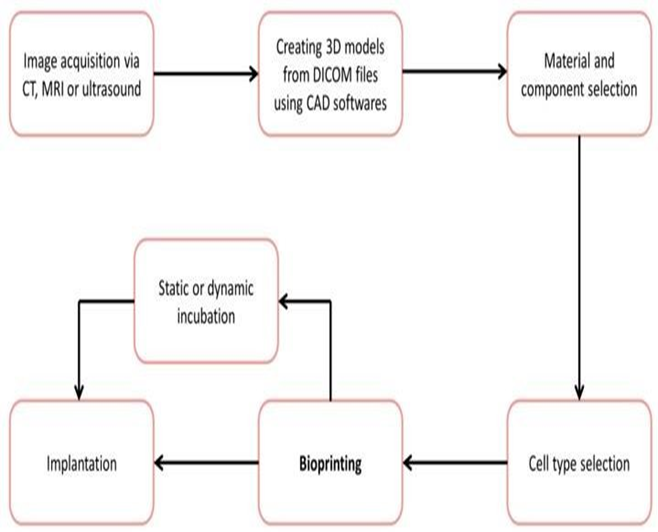
The process principally involves preparation, printing, maturation, and application. This can be summarized in the three key steps:
Pre-bioprinting:
Pre-bioprinting involves creating the digital model that the printer will produce. The technologies used are computed tomography (CT) and magnetic resonance imaging (MRI) scans.
Bioprinting:
Bioprinting is the actual printing process. Bio-ink is placed in a printer cartridge and deposition takes place based on the digital model.
Post-bioprinting:
Post-bioprinting is the mechanical and chemical stimulation of printed parts so as to create stable structures for the biological material.

- Basic Principle and Procedure of Fungal Staining
- What are Oxygen absorbers and How works?
- What is Pasteurization?
- What is Disinfectant and antiseptic? Disinfectants vs antiseptic
- Why More Men are Dying from Coronavirus than Women?
HOW DOES BIOPRINTING WORK?
Several bioprinting methods exist, based on either extrusion, inkjet, acoustic, or laser technologies. Despite the various types, a typical bioprinting process has a more- or-fewer standard series of steps:
- 3D Imaging: To get the exact dimensions of the tissue, a standard CT or MRI scan is used. 3D imaging should provide a perfect fit of the tissue with little or no adjustment required on the part of the surgeon.
- 3D Modelling: A blueprint is generated using AutoCAD software. The blueprint also includes layer- by-layer instruction in high detail. Fine adjustments may be made at this stage to avoid the transfer of defects.
- Bio ink Preparation: Bio ink is a combination of living cells and a compatible base, like collagen, gelatine, hyaluronan, silk, alginate or nanocellulose. The latter provides cells with scaffolding to grow on and nutriment to survive on. The complete substance is based on the patient and is function specific.
- Printing: The 3D printing process involves depositing the bio ink layer-by-layer, where each layer has a thickness of 0.5 mm or less. The delivery of smaller or larger deposits highly depends on the number of nozzles and the kind of tissue being printed. The mixture comes out of the nozzle as a highly viscous fluid.
- Solidification: As deposition takes place, the layer starts as a viscous liquid and solidifies to hold its shape. This happens as more layers are continuously deposited. The process of blending and solidification is known as crosslinking and may be aided by UV light, specific chemicals, or heat (also typically delivered via a UV light source).
VARIOUS TECHNIQUES:
1. INKJET-BASED 3D BIOPRINTING:
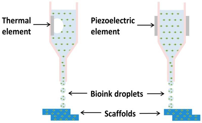
Inkjet bioprinting allows for the precise positioning of cells, with some studies achieving as few as a singular cell per printed droplet. Cells and biomaterials are patterned into a desired pattern using droplets, ejected via thermal or piezoelectric processes. Inkjet bioprinting is of great interest as it exhibits high resolution and cell viability. With this process, the accurate position of multiple cell types is possible. However, the limitations of vertical printing and restricted viscosities may mean that inkjet bioprinting needs to be combined with other printing techniques for future developments.
2. LASER-BASED BIOPRINTING:
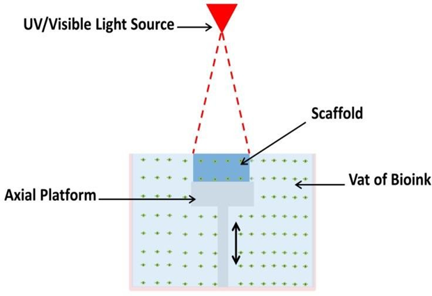
Stereolithography (SLA) is a technique that uses ultraviolet (UV) or visible light to cure photosensitive polymers in a layer-by-layer fashion. This nozzle-free technique eliminates the negative effects of shear pressure encountered when using nozzle-based bioprinting. It offers a fast and accurate fabrication, with resolutions ranging between 5–300 µm. Polymerization occurs at the top of the bio-ink vat where the biomaterial is exposed to light energy. After each layer is polymerized, the platform supporting the structure will be lowered in the vat, enabling a new layer to be photopolymerized on top.
3. LASER-BASED BIOPRINTING:
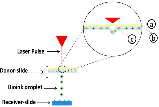
Laser-assisted printing was initially developed to deposit metals onto receiver sheets. Laser-assisted bioprinting (LAB) consists of three parts: a donor-slide (or ribbon), a laser pulse, and a receiver-slide. A ribbon is made of a layer of transparent glass, a thin layer of metal, and a layer of bio-ink. The bio-ink is transferred from the ribbon onto the receiver slide when the metal layer under the hydrogel is vaporized by a laser pulse. This scaffold-free technique has very high cell viabilities (>95) and a resolution between 10–50 µm. Some studies using LAB have demonstrated an accuracy of a singular cell per droplet. LAB has the ability to position multiple cell types with a high degree of accuracy, with several studies demonstrating singular the capability of positioning a singular cell per droplet. However, it is an expensive process to perform and suffers from low stability and scalability. It has shown great potential when combined with other bio-fabrication techniques.
4. EXTRUSION-BASED BIOPRINTING:
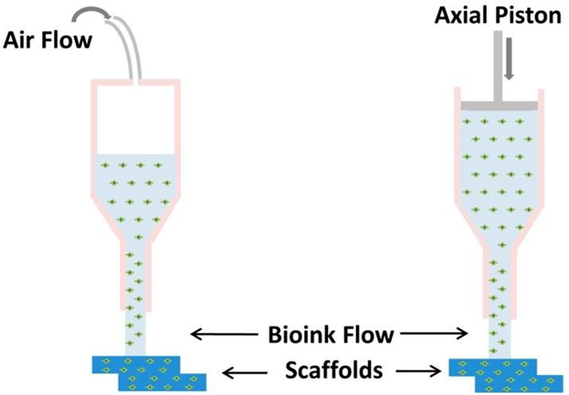
Extrusion-based printing is a pressure-driven technology. The bio-ink is extruded through a nozzle, driven either by pneumatic or mechanical pressure and deposited in a predesigned structure. The main advantage of extrusion bioprinting is the ability to print with very high cell densities. Despite its versatility and benefits, it has some disadvantages when compared to other technologies. The resolution is very limited, as a minimum feature size is generally over 100 µm, which is a poorer resolution than that of other bioprinting techniques. Extrusion printing can be regarded as a promising technology that allows the fabrication of organized constructs at clinically relevant sizes within a reasonable time frame. However, the selection of biomaterial and bio-ink concentration is important for the survival of the cells during fabrication. Also the maintenance of cell viability and functionality post-printing.
WHY IS 3D BIOPRINTING IMPORTANT?
The greatest importance of bioprinting lies in the resulting tissue-like structures that mimic the actual micro-and macro-environment of human tissues and organs. This is critical in drug testing and clinical trials, with the potential, for example, to drastically reduce the need for animal trials.
When living tissues and organs need not come from humans, this budding technology offers other massive opportunities. One example is testing treatment for diseases using artificially affected tissues.
The process could also eradicate the headaches associated with organ donation and transplantation. Apart from the lack of available organs, the entire process is criticized from a moral and ethical perspective. Organ replacement is the main objective, but tissue repair is also possible in the meantime. With bio-ink, it’s much easier to solve problems on a patient-specific level, promoting simpler operations.
APPLICATIONS of 3D Bioprinting:
Few of the main application areas of bioprinting:
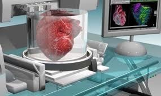
- Artificial organs are one of the greatest drivers of the technology due to the high rise of vital organ failure. Availability of 3D printed organs helps to solve organ-related issues faster and quicker. It is important to patients, their families, and healthcare systems.
- Development of tissues for pharmaceutical testing, when 3D printed, is a more cost- effective and ethical option. It also helps in identifying side effects of drugs. It also allows recommended drugs to be administered to humans with validated safe dosages.
- Cosmetic surgery, particularly plastic surgery and skin grafting also benefits from the technology. In this particular application, bio printed skin tissue could be commercialized. Some 3D printed tissues are already being bio printed for research on therapeutic purposes.
- Bone tissue regeneration as well as prosthetics and dental applications.
- There are various other uses and applications of bioprinting, including producing foodstuffs such as meat and vegetables
CONCLUSION:
Additive manufacturing has been heavily applied to tissue engineering over the past decade. Bioprinting enables the production of scaffolds with a homogeneous distribution of cells throughout a scaffold. Organized distribution of different cell types can be positioned within the supporting material, mimicking tissues with multiple cell types or the interface between two tissues. While the choice of material and design impact the viability and proliferation of the printed cells, the different techniques have also shown variable cell activities post-fabrication. Bioprinting is still under development and has many bridges to cross before entering the clinical world, particularly as an in situ direct application. From this brief review, it is concluded that different applications require different fabrication techniques, depending on required resolution, speed, cost, the ability to print vertically, etc. Future developments are now concentrating on the combining of techniques to work in a complementary fashion to optimize the process of creating tissue-mimicking structures.

- What are Windows Security Policies or group policy management for the GxP computerized system?
- How to write User Requirement Specification?
- Ransh Pharma: Best Intermediates Manufacturing Unit at Visakhapatnam
- What is 3D Bioprinting? Basic Principles, Techniques, and Application of 3D Bioprinting.
- Basic Principle and Procedure of Fungal Staining
- OXYGEN MANAGEMENT FOR SUSTAINABLE ENVIRONMENT
- mRNA VACCINES FOR COVID-19
- LITHIUM-ION BATTERIES An idea towards energy sustainability
- Artificial Leaf – A Blessing for Energy Crisis
- What are Oxygen absorbers and How works?
- What is Pasteurization?
- What is Disinfectant and antiseptic? Disinfectants vs antiseptic
- Why More Men are Dying from Coronavirus than Women?
- HOW COVID-19 SPREADS?
- What are the COVID-19 Symptoms?
- What is Coronavirus? How Coronavirus can Enter into our Body?
- Telemedicine Industry Analysis of Key Trends and Drivers Shaping Future Growth
- Rising Prevalence of Adverse Drug Reactions Drives Global Market for Pharmacovigilance
- Common Pharma Abbreviations
- Bioremediation : Promise for Eco-friendly enact|What is Bioremediation?
- GENERAL PROCEDURE FOR PASSIVATION
- How the New Drug approval process of CDSCO works?
- What is Passivation? How Does Passivation Process Work? How to Passivate Stainless Steel Parts?
- Explore new openings in Pharma Profession by Making yourself Skilled as Technical Associate in The Central Drugs Standard Control Organisation CDSCO
- What is Data Integrity and ALCOA Plus
- GOWNING PROCEDURE IN CLEAN ROOM
- How to Crack Your Job Interview?